Deep muscles · Lesson 7
Rotator cuff exercises: should you go light or heavy?
Light. On external rotations, in 18 men, the cuff, measured at the infraspinatus, and the rear deltoid both worked harder as the load went up, but the deltoid’s share grew: the authors recommend a low to medium load. Another team puts the sweet spot between 10 and 40% of your maximum force.
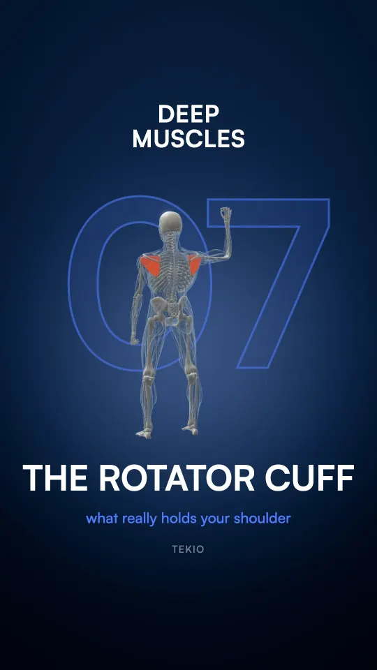
What is the rotator cuff?
It’s four muscles of your shoulder: the supraspinatus, infraspinatus, teres minor and subscapularis (Sharkey and Marder, 1995). In 32 dissected cuffs, the tendons of the subscapularis, supraspinatus and infraspinatus splayed out and interwove into a single, continuous insertion on the head of your humerus, your upper arm bone: that’s the “cuff” (Clark and Harryman, 1992).
The infraspinatus and teres minor turn your arm outward: that’s external rotation. In 10 adults, electrodes placed in these muscles found them most active in side-lying external rotation, out of 7 exercises (Reinold et al., 2004).
What does the rotator cuff do?
It holds your shoulder in place: its muscles stabilize it by pressing the head of the humerus into its socket, the glenoid (Parsons et al., 2002).
And your shoulder needs it: among a million insured people followed for six years in Taiwan, it was the joint that dislocated most, twice as often as the elbow (Yang et al., 2011). In US emergency rooms, it’s also the most common sports dislocation (Meixner and Loder, 2020).
Why does your shoulder need its cuff?
Because its socket is small. In 93 cadaver shoulders with no sign of disease, the head of the humerus was nearly always more curved than its flatter socket, which covers only a small part of it (McPherson et al., 1997). On CT scans of 239 patients treated for a fracture of the socket’s rim, its diameter was about two thirds of the head’s (Gordins et al., 2025).
What holds it is being pressed in: in 10 cadaver shoulders, the pressed-in head resisted pushes of up to 60% of the force pressing it. According to the authors, this mechanism may be important in the middle of a movement, where the capsule and ligaments are slack (Lippitt et al., 1993).
What does the cuff do when you raise your arm?
Your deltoid pulls your humerus up, your cuff pulls its head down. In 5 cadaver shoulders where a machine simulated muscle contraction, the deltoid alone made the head of the humerus rise as the arm lifted; with the whole cuff, it stayed centered at 30° and rose only 1.5 mm by 120° (Sharkey and Marder, 1995).
In 20 men without shoulder problems, filmed with X-rays raising their arm before and after their cuff was tired out, the head of the humerus rose more after fatigue (Teyhen et al., 2008). In 12 men, after exercises that tired the deltoid and the cuff, it moved 2.5 mm on average between arm positions, versus 0.3 mm at most before (Chen et al., 1999); it also rose in 20 adults when the nerve to the supraspinatus and infraspinatus was numbed (San Juan et al., 2013).
Light or heavy to train the rotator cuff?
In 18 men, electrodes measured the infraspinatus, a cuff muscle, and the rear deltoid during four external rotations at low, medium and high load. Both worked harder as the load rose, but the infraspinatus’s share dropped: the authors recommend a low to medium load to target it while limiting the deltoid, standing or lying face down (Kim et al., 2025).
Another team found the infraspinatus’s biggest share between 10 and 40% of maximum force, in 18 adults doing static external rotation (Bitter et al., 2007).
These are electrodes: activity, not growth. No study has yet measured which exercise makes the rotator cuff grow.
Your workout plan, built for your body.
In your Tekio session, on external rotations, go light then.

Sources
Studies checked in PubMed. Measured muscle activity isn’t growth: each fact keeps its study’s population and limits.
- Kim TG, Ma R, Yu IY (2025). A study of effective exercise methods and resistance intensity for selective strengthening of the infraspinatus muscle. J Back Musculoskelet Rehabil 38(2):304-313. PubMed · DOI
- Bitter NL, Clisby EF, Jones MA, et al. (2007). Relative contributions of infraspinatus and deltoid during external rotation in healthy shoulders. J Shoulder Elbow Surg 16(5):563-8. PubMed · DOI
- Reinold MM, Wilk KE, Fleisig GS, et al. (2004). Electromyographic analysis of the rotator cuff and deltoid musculature during common shoulder external rotation exercises. J Orthop Sports Phys Ther 34(7):385-94. PubMed · DOI
- Clark JM, Harryman DT (1992). Tendons, ligaments, and capsule of the rotator cuff. Gross and microscopic anatomy. J Bone Joint Surg Am 74(5):713-25. PubMed
- Sharkey NA, Marder RA (1995). The rotator cuff opposes superior translation of the humeral head. Am J Sports Med 23(3):270-5. PubMed · DOI
- Parsons IM, Apreleva M, Fu FH, et al. (2002). The effect of rotator cuff tears on reaction forces at the glenohumeral joint. J Orthop Res 20(3):439-46. PubMed · DOI
- Yang NP, Chen HC, Phan DV, et al. (2011). Epidemiological survey of orthopedic joint dislocations based on nationwide insurance data in Taiwan, 2000-2005. BMC Musculoskelet Disord 12:253. PubMed · DOI
- Meixner C, Loder RT (2020). The Demographics of Fractures and Dislocations Across the Entire United States due to Common Sports and Recreational Activities. Sports Health 12(2):159-169. PubMed · DOI
- McPherson EJ, Friedman RJ, An YH, et al. (1997). Anthropometric study of normal glenohumeral relationships. J Shoulder Elbow Surg 6(2):105-12. PubMed · DOI
- Gordins V, Sansone M, Karlsson L, et al. (2025). Bony Bankart lesions exceeding 20% of glenoid width are common and show gender-based anatomical differences: A radiological study of 239 patients. J ISAKOS 15:101001. PubMed · DOI
- Lippitt SB, Vanderhooft JE, Harris SL, et al. (1993). Glenohumeral stability from concavity-compression: A quantitative analysis. J Shoulder Elbow Surg 2(1):27-35. PubMed · DOI
- Teyhen DS, Miller JM, Middag TR, et al. (2008). Rotator cuff fatigue and glenohumeral kinematics in participants without shoulder dysfunction. J Athl Train 43(4):352-8. PubMed · DOI
- Chen SK, Simonian PT, Wickiewicz TL, et al. (1999). Radiographic evaluation of glenohumeral kinematics: a muscle fatigue model. J Shoulder Elbow Surg 8(1):49-52. PubMed · DOI
- San Juan JG, Kosek P, Karduna AR (2013). Humeral head translation after a suprascapular nerve block. J Appl Biomech 29(4):371-9. PubMed · DOI
These lessons inform; they don’t replace advice from a health professional.